Options For Diabetic Retinopathy Screenings
Now that you’ve used the Retina Risk app to assess your risk of diabetic retinopathy (DR), there are several different methods of eye screening to consider. Depending on your location, different or limited screening options may be accessible to you. Always speak to a healthcare professional about your options and which one is right for you.
Retinopathy screening programs
Retinopathy screening programs were introduced in the early 1990s which succeeded in reducing cases of vision loss due to DR. As cases of diabetes and related vision loss continue to rise, new technology is being developed, that will make eye screening faster, less expensive and more convenient for the patient. This will help prevent eyesight deterioration from the very beginning, as early detection is the most significant factor in therapeutic success.
Dilated fundus examination
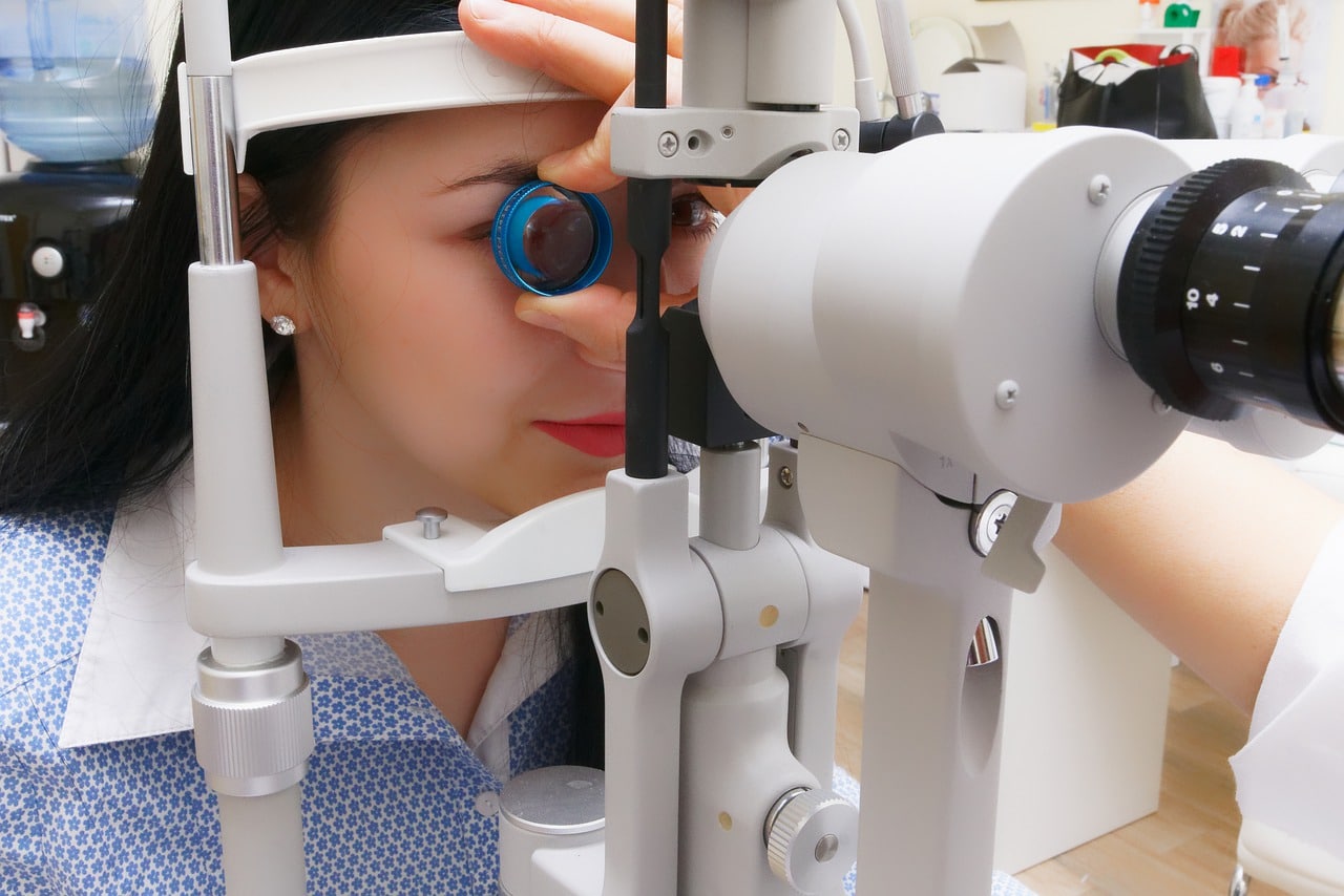
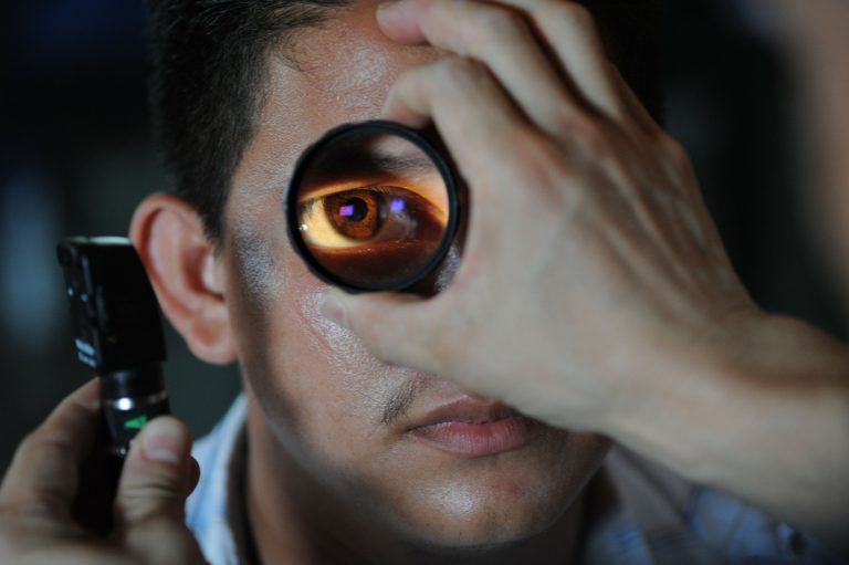
The most common and cost-effective screening method for DR is the dilated fundus examination. Eye drops are used to dilate the pupil so that the ophthalmologist can examine the back of the eye. Sometimes a fundus photograph is taken, so that the image can be analyzed further. Non-dilation fundus photography is also an option; however, dilating the pupil reduces the chance of having ungradable photographs from 19-26% to 4-5%.
Fluorescein angiography
A different type of procedure is fluorescein angiography. A fluorescent dye is injected into the patient’s bloodstream. This highlights the blood vessels in the back of the eye so that it can be photographed.
This form of examination is an accurate method to determine whether there is adequate blood flow through the blood vessels in the back of your eye. It can also detect vascular changes due to the inner and outer blood-retinal barrier.
Fluorescein angiography can be helpful in observing whether diabetic retinopathy treatment is working. This method of screening can be problematic as it’s an invasive procedure, but at the same time highly effective at detecting any issues in the eye.
Optical coherence tomography (OCT)
As technology evolves, better cameras and machines become available to help increase early diagnosis in diabetic retinopathy. One such development has been the optical coherence tomography (OCT) imaging technique. Thanks to high-speed scanning and sensitivity improvements, screening with OCT allows for a 3-D high-quality image of the eye.
OCT screening is a cost-effective way to screen for diabetic retinopathy, but it does come with high implementation costs for the clinic.
Recently, the Lions Club of Iceland donated two OCT cameras to the University Hospital of Iceland and the Service Center for the Visually Impaired, greatly increasing the chances of patients getting a faster and more reliable non-invasive screening.
Future of diabetic retinopathy screening
The future of DR screening is focused on creating options that are more accessible to everyone. Automated DR image assessment systems are in development, which will alleviate pressure on doctors to analyse images from DR screening, meaning that more patients can be handled.
Less invasive techniques will also be the norm: OCT angiography will be a noninvasive screening technique that can detect blood flow from within the retina without the need to inject patients with fluorescent dye.
Smartphones for portable eye screening
Smartphones will also play a big role in DR screening. Modern smartphone cameras are already producing images that have better quality that clinical retinal cameras. Using smartphones and a specialised handheld lens, DR screening can become low cost and portable. This will make it a viable option in countries where more expensive machinery may be unavailable

