Does vitrectomy improve your vision?
Vitrectomy is a type of eye surgery that is performed to treat various problems with the retina and vitreous. It can often improve or stabilize vision, but to what extent depends on various factors, including the health of the eye before the surgery. The healthier the eye is before surgery, the greater hope you can have for vision improvement and quick healing.
In a recent study, researchers sought to determine the visual gain following a vitrectomy for diabetic tractional retinal detachment. This condition, in the setting of proliferative diabetic retinopathy, is one of the most advanced stages of diabetic retinopathy. The researchers concluded that before surgery nearly 80% of the eyes had low vision. Following vitrectomy, over half of these eyes had significant improvements in visual acuity.
In another large retrospective study of patients with advanced diabetic tractional retinal detachment, researchers found that vitrectomy achieved excellent anatomical outcome and improved or stabilized vision in 80.1% of eyes.
Your vision may not be completely normal immediately following your vitrectomy. This is , especially true if your condition caused permanent damage to your retina. Some patients seem to experience a decrease in vision for the first few days following the procedure. Others may need weeks and sometimes months before feeling significant improvement in vision.
It is important that you ask your eye doctor about how much improvement you can expect and how long the healing may take for your eyes.
What is vitrectomy?
Vitrectomy involves the removal of the vitreous humor, which is a transparent gel-like substance that forms nearly 80 percent of the eyeball. The vitreous humor provides a medium through which light can travel inside the eye to reach the retina.
During vitrectomy, your surgeon removes some or all of the vitreous from the middle of your eye. This allows for a variety of repairs, including removal of scar tissue, laser repair of retinal detachments and treatment of macular holes.
A vitrectomy is sometimes performed even in patients with a normal vitreous humor, so that the ophthalmologist surgeon can access the retinal tissues properly to repair a tear or hole in the retina.
Once the surgery is complete, the vitreous is replaced with either a salt water (saline) solution or a bubble made of gas or oil. This helps to hold the retina in position.
Below you´ll find more details about what vitrectomy entails, when it may be required, and what happens before, during, and after the surgery.
How long does it take for clear vision after a vitrectomy?
In most cases, it takes around 2 to 4 weeks for the vision to become clear after the vitrectomy.
The extent of the clarity of the eyesight after the surgery depends on several factors, including:
- During vitrectomy, multiple incisions may be taken on the white of the eye called the sclera. If the stitches are close to the cornea, they may change the shape of the cornea resulting in blurry vision.
- The eye drops used for the dilatation of the eyes for the surgery may cause blurring of the eyesight. Your doctor may recommend that you continue to use these drops for 5 to 7 days after the procedure. This may cause the blurry vision to persist for longer.
- If the vitrectomy is performed to repair a larger hole in the retina, the retinal tissues may not heal completely, possibly resulting in persistent vision loss.
- If the vitrectomy is performed to repair the swelling in the retina caused by diabetes, it may heal to some extent, causing the eyesight to be clearer. This is dependent on good blood sugar control with appropriate treatment and possibly a change in diet or lifestyle.

When do you require a Vitrectomy?
Your ophthalmologist may recommend a vitrectomy if you have for example any of these diseases or conditions:
- Diabetic retinopathy, with bleeding or scar tissue affecting the retina or vitreous gel.
- Some forms of retinal detachment, which happens when the retina lifts away from the back of the eye.
- Macular hole which refers to a tear or opening in your macula.
- Macular pucker, which happens when wrinkles, creases or bulges form on your macula.
- Endophthalmitis, which is an infection of the tissues or fluids inside the eyeball. It may be caused by severe infections and swelling of the vitreous and other parts of the inside of the eye.
- Severe eye injury.
- Vitreous hemorrhage (abnormal bleeding inside the vitreous)
How do I prepare for a vitrectomy?
You will prepare for a vitrectomy in close consultation with your doctor and healthcare providers. There are few things you may want to ask your doctor specifically about in preparation for the vitrectomy surgery.
You need to find out whether you should stop taking any medicines before the procedure. If you are using any medicines that can affect the bleeding and clotting processes, you might have to stop using them for about 2 weeks before the surgery.
Your doctor may examine the retina using special instruments to shine a light in your eye. An ultrasound of the eye may be advised to assess the structural changes that have occurred in the retina.
In most cases, your doctor will require that you avoid eating anything after midnight before your surgery.
What Happens During The Vitrectomy?
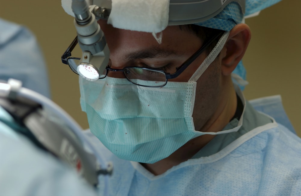
Vitrectomy is generally performed by a doctor specially trained in eye surgery. Discuss with your doctor the specificities of your operation, including what to expect, how long it will take and expected recovery.
Vitrectomy is often done in an outpatient surgery center. You will have a local or a general anesthesia to numb the eye. If your vitrectomy is performed under local anesthesia, this means that you´re awake during the surgery. Your doctor may use anesthetic eye drops or injections to ensure you don’t feel anything. In some cases, the procedure is performed under general anesthesia. Then you will sleep through the surgery.
The surgeon will make an incision in the outer layer of your eye. This is followed by a small incision in the sclera, which is the white part of the eye. Then, the vitreous is removed along with the scar tissues and foreign materials. At this point, your doctor will do any repairs to your eye as needed. This may include using a laser to fix a tear in your retina.
As mentioned before, your doctor will replace the vitreous with another sort of fluid to help keep the retina in place. The surgeon may need to close the incisions with stitches, though in some cases, stitches are not needed. Many vitrectomy procedures are now performed with self-sealing, sutureless (no-stitch) incisions approximately one half of a millimeter in size, which is about the width of an eyelash.
An antibiotic ointment will be placed on your eye to help prevent infection and your eye will be covered with a patch. The surgery can take from one to several hours.

What Happens After a Vitrectomy Procedure?
In most cases, patients are able to go home within a few hours following a vitrectomy. You will be monitored as you rest and recover from anesthesia. Then you can go home. As you´ll not be able to drive, you´ll need to make alternative arrangements.
Following the surgery it’s important to comply with your doctor’s instructions about eye care. Your ophthalmologist may prescribe medicine to help the eye heal. You may need to take eye drops with antibiotics to help prevent infection. You may also need to wear an eye patch for a day or so to protect the eye.
Pain is rather rare after vitrectomy. Some patients feel a scratchy, sandy, or gritty sensation, as if something is inside the eye. This will disappear with the medicines and time. If you feel any soreness in the eye that persists for a few days, it can be relieved by taking over-the-counter pain relievers.
If a gas bubble was placed in your eye, you´ll be told to refrain from air travel for a while after the surgery. This is because rapid altitude changes can affect the size of the bubble. Your doctor will inform you when you can safely return to your normal activities.
Conclusion
Although vitrectomy improves or stabilizes vision in most cases, vision may not be fully normal after the surgery. This is especially true if your condition caused permanent damage to your retina. Vitrectomy is generally a safe procedure. It should be performed by a skilled and experienced ophthalmic surgeon to ensure successful outcomes without the risk of adverse effects. Bear in mind that diabetic retinopathy can be prevented if diagnosed early!
You need to follow-up with your doctor to see whether the procedure was effective. It is crucial that you notify your doctor immediately if you experience increasing pain or decreased vision.

